Piattaforma di Imaging Biomedico e Radiomica
Description
During 2021, the Biomedical Image Processing, Radiomics and Analysis platform utilized two magnetic resonance systems (3T and 7T), a Spectrum In Vivo imaging system (Bioluminescence), and Positron Emission Tomography – Computed Tomography (PET/CT) made available by the participating institutions. Pre-processing, segmentation, radiomics, and artificial intelligence (machine learning and deep learning) tools were developed from imaging data for predictive diagnosis of diseases and medical diagnostic support, in collaboration with GIT and IBFM-CNR. The platform provides crucial support to foster the translation of scientific results into clinical applications, particularly in the fields of neuroscience and cancer research. The staff, currently expanding, consists of a computer scientist specialized in biomedical image processing and analysis and in clinical and preclinical magnetic resonance acquisition, a physicist, a veterinarian, and a PhD student in nuclear medicine. During 2022, the platform will be enhanced with a WIZARD 2470 gamma counter with 10 PerkinElmer detectors and radioprotection devices (at ISMETT), a Skyscan 1276 CMOS microCT Bruker, and a Magnetic Resonance guided Focused UltraSound Surgery (MRgFUS) system – ExAblate 2100 – InSightec (at IZS), in order to provide additional imaging and radiomics capabilities.
Expertise
- Image processing models (MRI / PET / CT / IVIS and histological), 3D segmentation, deep learning and machine learning to extract, classify, and delineate tumor volumes and radiomic features for predictive diagnosis of diseases (e.g., tumor, COVID-19), recurrence assessment, and medical decision support • Magnetic Resonance Imaging (T1, T2, PD, DWI, ADC and DCE)
- Positron Emission Tomography / Computed Tomography (PET / CT)
- Spectroscopy on phantoms, in vivo and ex vivo samples
- Radiobiology studies in vitro and in vivo on innovative radiopharmaceuticals
- Magnetic Resonance-guided Focused Ultrasound Surgery in vitro and in vivo (MRgFUS)
Facilities & Equipment
Software
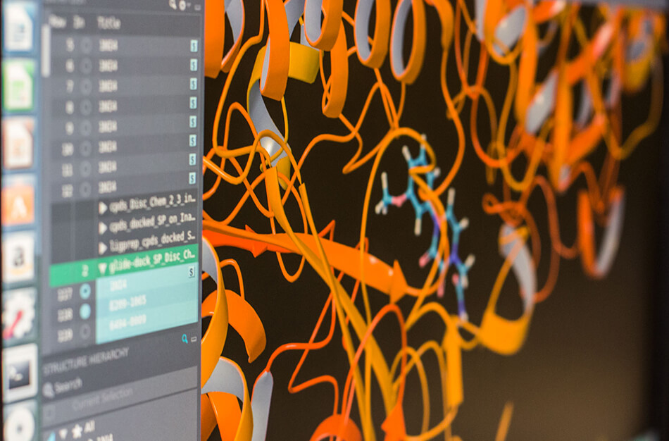
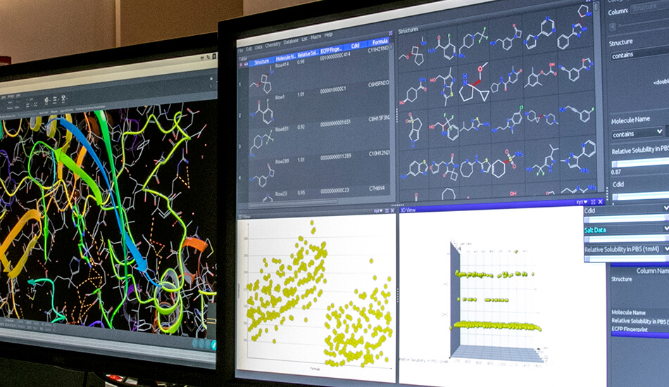
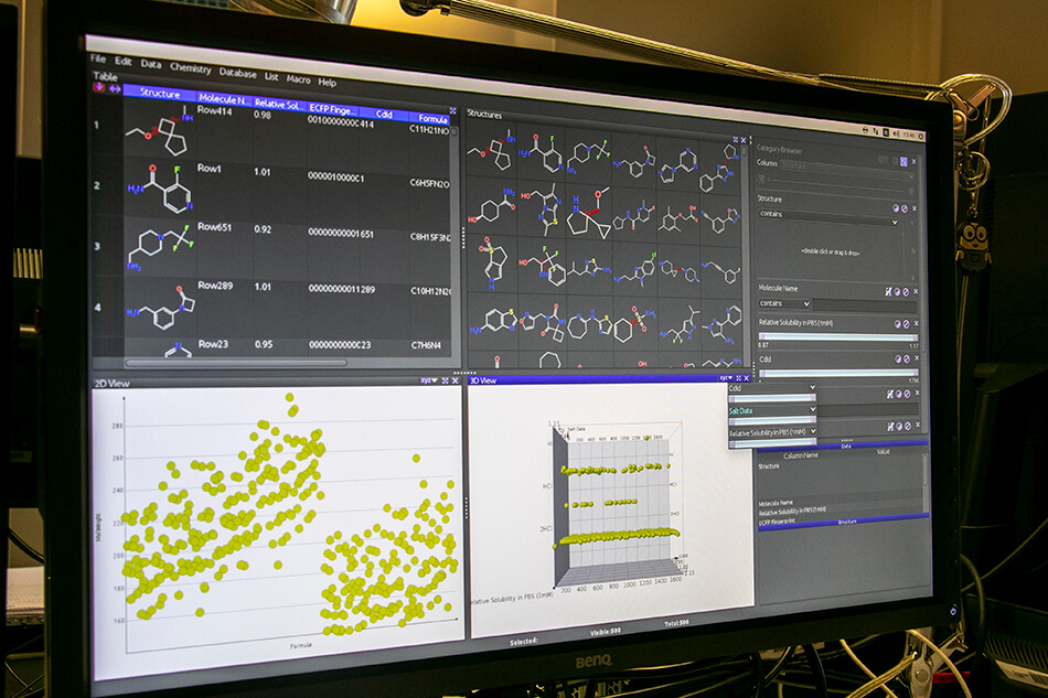
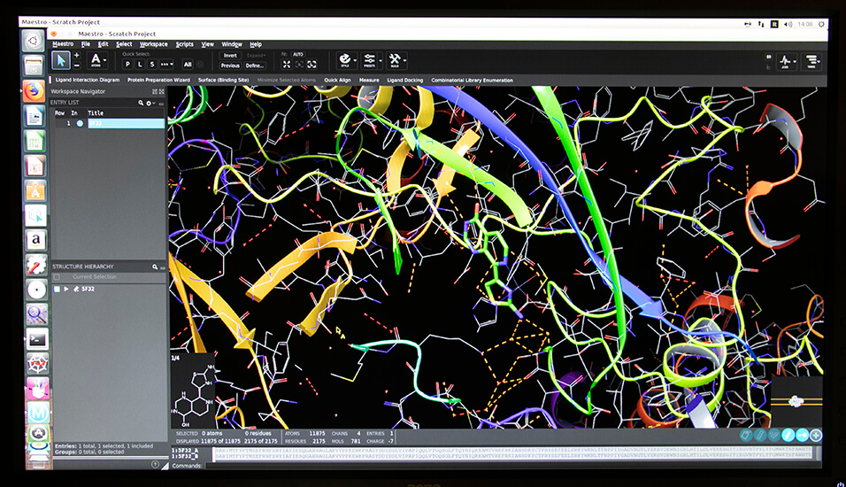
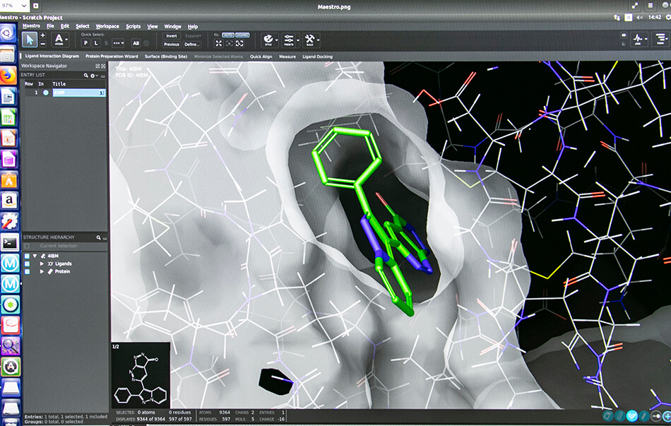
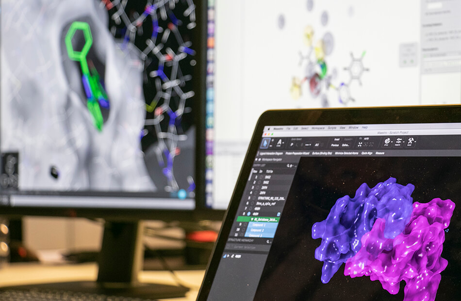
- Schrodinger suite for small molecule drug discovery;
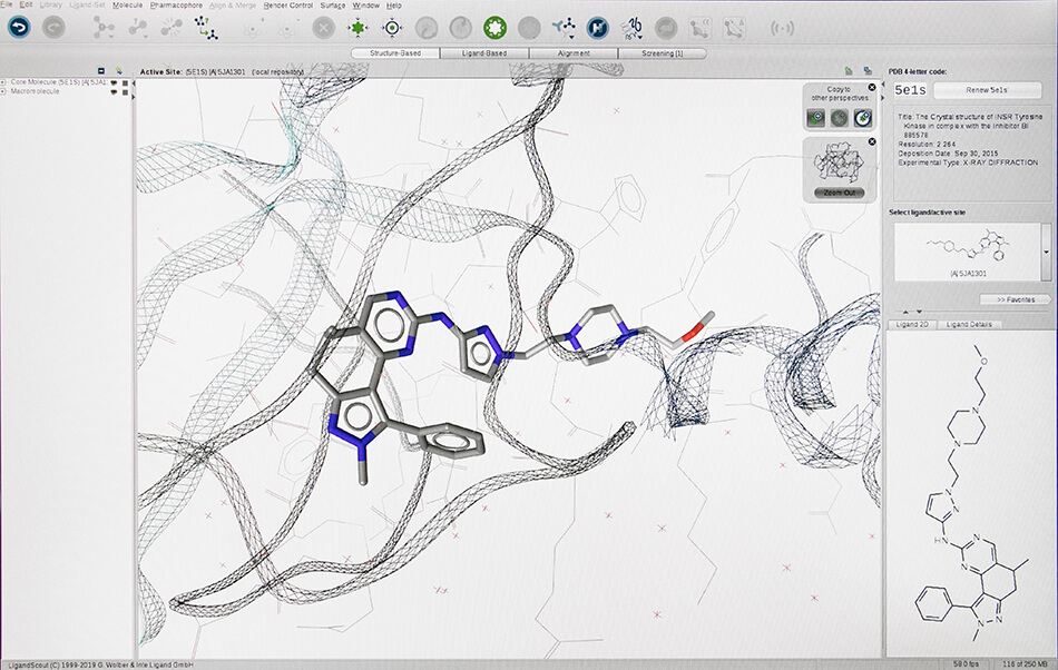
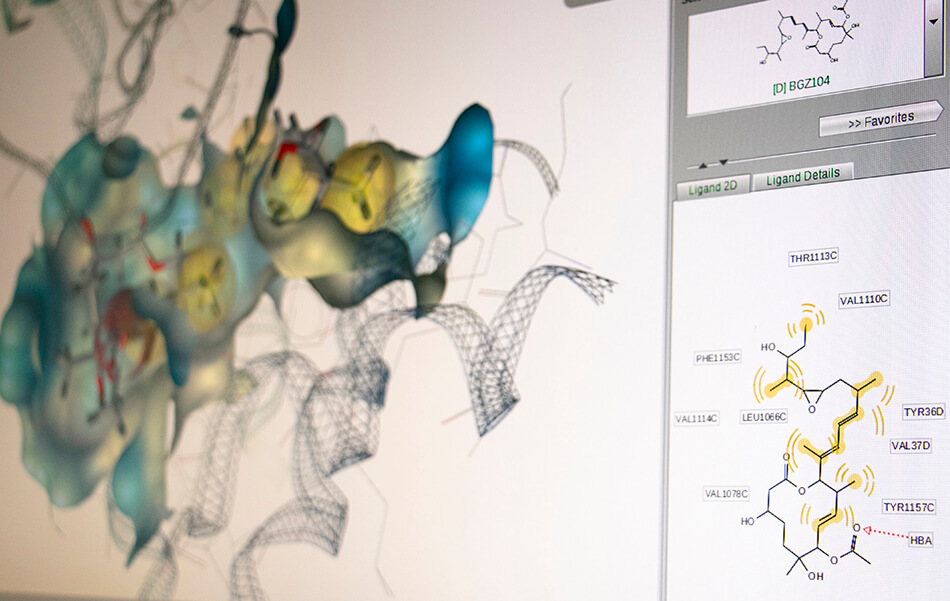
- LigandScout expert suite;
- Autodock and Autodock Vina;
- Desmond (OPLS2005 and OPLS3e);
- AMBER;
- NAMD;
- VMD;
- Gromacs;
- RDKit;
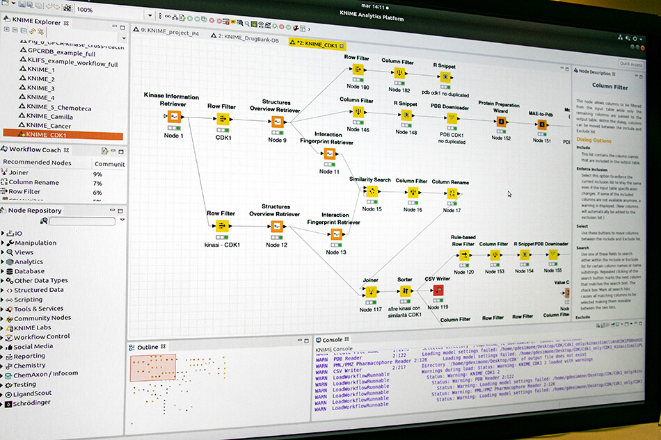
- KNIME.
Hardware
- 6 Workstations;
- Server in HPC mode: 200 CPUs and 2 x NVIDIA Tesla K80.
Computational capacity:
- Library optimisation → ~ 6,000 molecules/min
- Virtual Screening HTVS → ~ 5,000 molecules/min
- Virtual Screening SP → ~ 1,500 molecules/min
- Molecular Dynamics → ~ 200 ns/day/GPU (on an average system of 40,000 atoms)
Integrated in silico platform
The group is developing an integrated platform for the study of molecular networks in collaboration with the bioinformatics group.
Contacts:
Collaboration:
- IRCCS ISMETT, Palermo, Italia
- Istituto di Bioimmagini e Fisiologia Molecolare, (IBFM-CNR), Cefalù, Italia
- Georgia Institute of Technology, (GIT), Atlanta, USA
- Unità di Medicina Nucleare, Università di Messina, Italia
- Dipartimento di Promozione della Salute, Assistenza Madre e Infanzia, Medicina Interna e Specialità Mediche, (PROMISE), Università di Palermo, Italia
- Dipartimento di Biomedicina, Neuroscienze e Diagnostica avanzata (BIND), University of Palermo, Palermo, Italia
- Dipartimento di Ingegneria, Università di Palermo, Italia
- Dipartimento di Scienze Agrarie, Alimentari e Forestali
- Università degli Studi di Palermo Unità di Fisica Medica, Ospedale Cannizzaro, Catania, Italia
- Dipartimento di Medicina Nucleare, Ospedale Cannizzaro, Catania, Italia Istituto Zooprofilattico Sicilia (IZS), Palermo, Italia
Hardware
I am text block. Click edit button to change this text. Lorem ipsum dolor sit amet, consectetur adipiscing elit. Ut elit tellus, luctus nec ullamcorper mattis, pulvinar dapibus leo.