
Development of AI Algorithms for Extraction and Selection of Radiomics Features from Biomedical Imaging
Abstract
Radiomics is a new frontier of medicine based on the extraction of quantitative data from radiological images which can not be seen by radiologist’s naked eye and on the use of these data for the creation of clinical decision support systems. The long-term goal of radiomics is to improve the non-invasive diagnosis of focal and diffuse diseases of different organs by understanding links between extracted quantitative imaging data and the underlying molecular and pathological characteristics of lesions. In the last decade, several studies have highlighted the enormous potential of radiomics in both tumoral and non-tumoral diseases of many organs and systems including brain, lung, breast, gastrointestinal and genitourinary tracts. The enormous potential of radiomics needs to be pursued with the methodological rigor of scientific research and by integrating radiological data with other medical disciplines, in order to improve personalized patient management.
Impact:
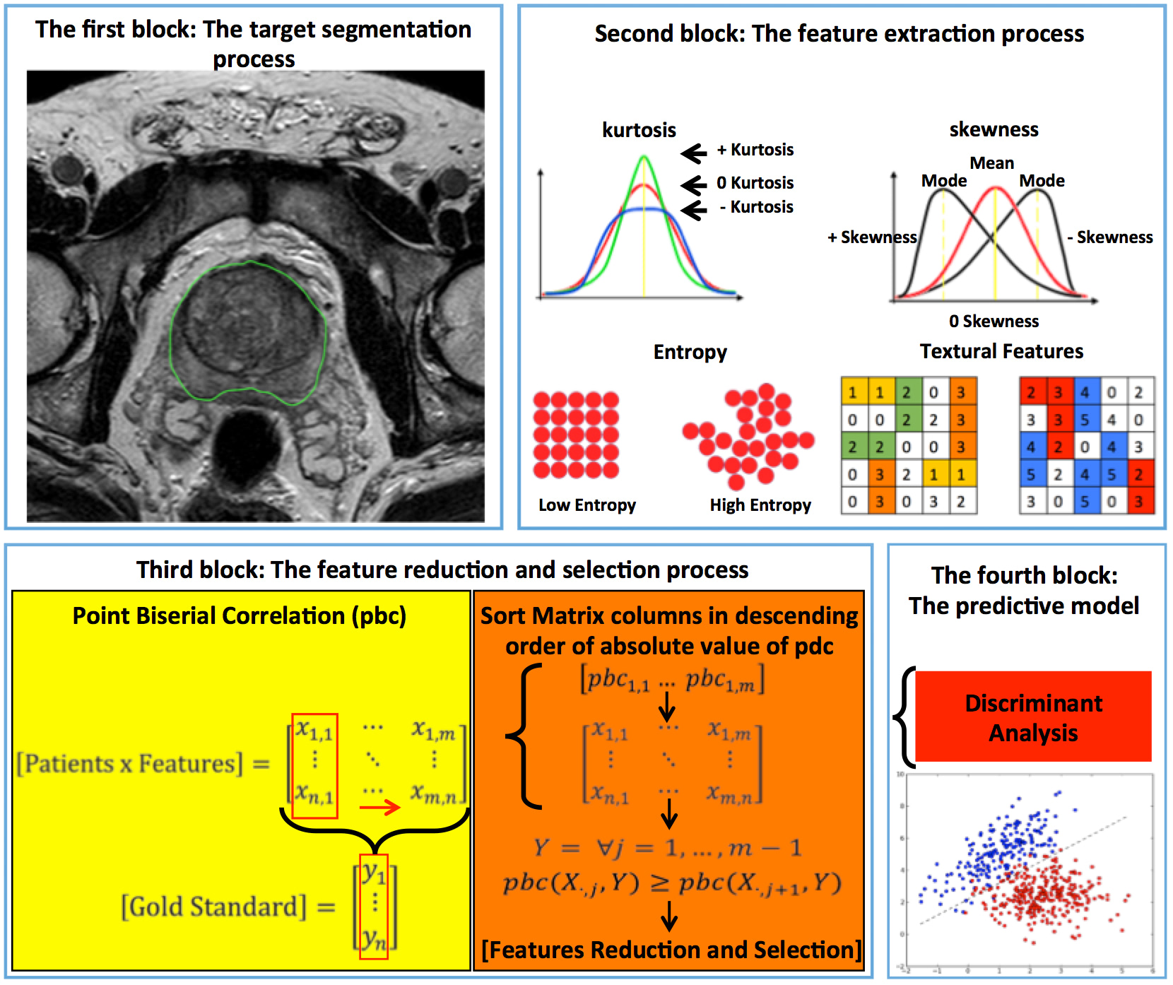
Radiomics is utilized to improve the prediction of patient overall survival and/or outcome, identifying personalized predictive and/or prognostic models to support the medical decision process. Target segmentation, feature extraction, feature selection, and classification model are the fundamental blocks of a radiomics workflow. The good diagnostic performance of the combination of multiple radiomic features for the diagnosis of cancer may help predicting lesions where aggressive management may be warranted. The project proposes a novel radiomics workflow to identify a relevant prognostic model concerning a real clinical problem. In the specific, we propose an operator-independent segmentation system with the consequent automatic extraction of radiomics features, and a novel feature selection approach to create a relevant predictive model in patients underwent different imaging methods like magnetic resonance, computer tomography and positron emission tomography.
Pipeline
-
CLINICAL
NEED -
DISEASES
ANALYSIS - DISCOVERY
-
PRECLINICAL
VALIDATION -
PRECLINICAL
DEVELOPMENT -
CLINICAL
STUDIES
Principal Investigator
Contact
Therapeutic Areas:
Product:
Biomarkers, Medical Devices & Tissue Engineering
Collaborations:
- Istituto Mediterraneo per i Trapianti (ISMETT) IRCCS, Palermo, Italia
- Istituto di Bioimmagini e Fisiologia Molecolare (IBFM-CNR), Cefalù, Italia
- Georgia Institute of Technology (GIT), Atlanta, USA
Unità di Medicina Nucleare, Università di Messina, Messina, Italia - Dipartimento di Biomedicina, Neuroscienze e Diagnostica avanzata (BIND), University of Palermo, Palermo, Italia
- Unità di Medicina Nucleare, Fondazione Istituto G.Giglio, Cefalù, Italia
- Dipartimento di Ingegneria, Università di Palermo, Palermo, Italia
- Unità di Fisica Medica, Ospedale Cannizzaro, Catania, Italia
Dipartimento di Medicina Nucleare, Ospedale Cannizzaro, Catania, Italia
Scarica il pdf del progetto